
Digital X-Rays — Advanced Imaging with 90% Less Radiation in Rockville, Bethesda & DC Metro
Experience safer, faster dental imaging with our advanced digital X-ray technology. Natural Dentist Associates provides comprehensive diagnostic imaging with minimal radiation exposure throughout Rockville, Bethesda, and the DC metro area.
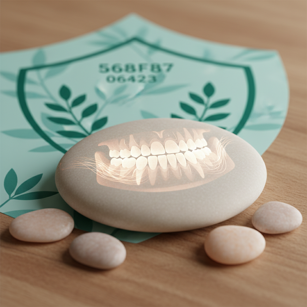
Digital radiography has revolutionized dental diagnosis, providing superior image quality with dramatically reduced radiation exposure. While traditional film X-rays remain useful, digital technology offers immediate viewing, enhanced diagnostic capability through image manipulation, easier sharing with specialists, and environmental benefits eliminating chemical processing.
At Natural Dentist Associates, we use the latest digital X-ray systems providing crystal-clear images with up to 90% less radiation than traditional film. Our commitment to biological dentistry means minimizing exposure while maximizing diagnostic accuracy for comprehensive, safe dental care.
Real experiences from patients who appreciate our advanced imaging technology
I was called in for my appointment right on time and my treatment was exemplary in every way. All the staff is friendly and accommodating and Dr. Brow…
I was called in for my appointment right on time and my treatment was exemplary in every way. All the staff is friendly and accommodating and Dr. Brown made me feel comfortable and secure. She took care of my needs quickly and gently. A very pleasant experience!
READ MOREDr. Baer is an excellent dentist. Accessible, helpful, does everything to make you comfortable and very professional. She does excellent work and …
Dr. Baer is an excellent dentist. Accessible, helpful, does everything to make you comfortable and very professional. She does excellent work and runs her practice efficiently and professionally.
READ MOREFantastic experience. Very professional
I am so grateful I learned about Dr. Baer from my chiropractor. My dental needs were not met by my prior general dentists , whose team only approached…
I am so grateful I learned about Dr. Baer from my chiropractor. My dental needs were not met by my prior general dentists , whose team only approached by dental care focused on six month cleanings, and no sign of cavities. Dr. Baer addressed my exam holistically, which is what I wanted but could not easily find from other dentists I was referred to. She diagnosed and treated my adult lip and tongue frenum constraints that in my generation were not recognized as they are today by this generation of pediatric dentists. I live in Northern Virginia but am happy to make the commute to their offices in Maryland for the best care. I am also working with a MYO Professional, Sarah, who has guided me through mouth exercises before and after my frenumectomies. I realize now how especially beneficial functional dentistry is, and I wish these dentists were more available and discoverable.
READ MOREDr Tipograph was outstanding in her presentation on what the procedure will entail and continued to assure me during the procedure . Her professiona…
Dr Tipograph was outstanding in her presentation on what the procedure will entail and continued to assure me during the procedure . Her professional and personal attitude made my visit enjoyable and pain free
READ MOREProfessional and personal. I feel like i belong there. They do TAKE CARE IF ME
Digital radiography offers numerous advantages over traditional film. Radiation exposure is reduced by 80-90% due to highly sensitive digital sensors requiring less exposure time. Image quality is superior with higher resolution, ability to adjust contrast and brightness revealing subtle details, and zoom capability examining specific areas. Efficiency improves dramatically with instant image viewing eliminating processing time, immediate availability for consultation, and easy electronic sharing with specialists or other dentists. Environmental impact is eliminated through no chemical processing or disposal, reduced waste, and paperless storage. Diagnostic capability is enhanced through measurement tools for precise treatment planning, side-by-side comparison of images over time, and enhancement features highlighting areas of concern. Storage and accessibility are simplified with secure digital archiving, no physical storage space required, and instant retrieval during appointments. While traditional film X-rays remain diagnostically useful, digital technology provides superior benefits in virtually every aspect of dental imaging supporting accurate diagnosis with minimal patient exposure.
Yes, digital X-rays are extremely safe with radiation exposure far below levels causing health concerns. Digital sensors require 80-90% less radiation than traditional film due to high sensitivity. A full mouth series of digital X-rays delivers approximately 0.005 mSv (millisieverts) of radiation—less than one day of natural background radiation from environment. For comparison, annual background radiation averages 3.0 mSv, a cross-country flight exposes you to 0.035 mSv, and a chest X-ray delivers about 0.1 mSv. Dental X-rays represent tiny fraction of radiation exposure encountered daily. Additional safety measures include lead aprons blocking scatter radiation, thyroid collars protecting sensitive thyroid gland, focused beam technology limiting radiation to target area, and digital sensors positioned precisely minimizing retakes. We follow ALARA principle (As Low As Reasonably Achievable) taking only necessary images based on clinical need. Our biological dentistry philosophy prioritizes minimizing exposure while maintaining diagnostic quality. For most patients, benefits of accurate diagnosis far outweigh minimal radiation risk. We’re happy to discuss concerns and explain why specific images are recommended.
X-ray frequency depends on individual oral health status, age, and risk factors. General guidelines for healthy adults include bitewing X-rays every 12-24 months checking for cavities between teeth and monitoring bone levels, and full mouth series or panoramic X-ray every 3-5 years for comprehensive evaluation. Higher risk patients may need more frequent imaging including every 6-12 months for history of cavities or gum disease, periodontal disease requiring monitoring, and orthodontic treatment needing regular assessment. New patients typically need baseline X-rays for comprehensive evaluation unless recent images are available from previous dentist. Children’s frequency varies based on cavity risk and development stage. We follow evidence-based guidelines from American Dental Association recommending X-rays based on individual needs rather than arbitrary schedules. Our biological approach means taking only images providing diagnostic value affecting treatment decisions. We never take X-rays simply because certain time has passed. During examination we assess whether X-rays would reveal information changing diagnosis or treatment plan. If not, we defer imaging until clinically indicated. This conservative evidence-based approach ensures you receive necessary imaging while minimizing exposure.
X-rays reveal problems invisible to clinical examination alone. Between teeth, X-rays show cavities forming on contact surfaces where teeth touch—areas impossible to see visually and representing 30-40% of all cavities. Below gumline, imaging reveals bone loss from periodontal disease, abscesses or infections at tooth roots, impacted teeth including wisdom teeth, cysts or tumors in jaw bone, and root fractures. Within teeth, X-rays detect decay under existing fillings, root canal problems, internal resorption, and developmental abnormalities. Structural issues revealed include jaw joint (TMJ) problems, sinus abnormalities, bone density and quality, and anatomical variations affecting treatment. Many serious problems cause no symptoms in early stages when treatment is simplest and most conservative. By the time pain develops, problems often require extensive treatment. X-rays allow early detection preventing minor issues from becoming major problems. For example, small cavity caught on X-ray needs simple filling, while same cavity missed until causing pain may require root canal and crown. Regular appropriate imaging represents preventive investment protecting oral health while minimizing treatment needs and associated concerns.
Yes, you have the right to decline any recommended treatment including X-rays. However, this decision has important implications. Without X-rays, dentists cannot detect hidden problems between teeth, beneath existing fillings, in bone structure, or at tooth roots. This limits our ability to provide comprehensive diagnosis and treatment planning. If you decline recommended imaging, we document this decision and may ask you to sign informed refusal acknowledging you understand risks of forgoing diagnosis. These risks include undetected cavities progressing to larger problems, periodontal disease advancing unchecked, infections developing without symptoms, and treatment performed without complete information. Some procedures cannot be safely or effectively performed without X-rays including root canal treatment requiring precise measurements, implant placement needing bone assessment, and orthodontic treatment requiring jaw relationship analysis. We respect your concerns and are happy to discuss why specific images are recommended, radiation exposure levels and safety measures, and alternatives if any exist. Our goal is informed decision-making where you understand both benefits of recommended imaging and risks of declining. Some patients prefer minimizing X-rays even with these limitations—we work within your preferences while ensuring you understand implications for diagnosis and treatment planning.
Dental X-rays during pregnancy are considered safe when properly performed with appropriate precautions, though we typically defer non-urgent imaging until after delivery when possible. The American College of Obstetricians and Gynecologists and American Dental Association state routine dental X-rays can be performed during pregnancy using proper shielding. Digital X-rays deliver minimal radiation with doses far below levels affecting fetal development. Radiation from full mouth series is less than one day of natural background exposure. With lead apron and thyroid collar, virtually no radiation reaches abdomen. However, we follow conservative approach deferring elective imaging until after pregnancy when possible. Emergency imaging for infection diagnosis or trauma evaluation may be necessary protecting both mother and baby from untreated dental infection. If X-rays are clinically necessary during pregnancy, we use extra precautions including double lead aprons, thyroid collars, minimal necessary images, and digital technology for lowest possible exposure. Untreated dental infections during pregnancy pose greater risk than properly performed X-rays. We work with your obstetrician coordinating care and discussing any concerns ensuring both dental and prenatal health are protected.
Patients throughout Rockville, Bethesda, Montgomery County, and Northern Virginia choose us for advanced digital X-ray technology combined with conservative biological dentistry protocols. Our convenient Rockville location off I-270 and I-495 provides easy access throughout the DC metro area. Our imaging approach includes state-of-the-art digital sensors providing 90% less radiation than film, instant high-resolution images with enhancement capabilities, comprehensive radiation protection with lead aprons and thyroid collars, evidence-based imaging taking only clinically necessary X-rays, and integration with 3D cone beam for complex cases. Our biological dentistry philosophy means minimizing radiation exposure while maintaining diagnostic accuracy. Many patients appreciate our conservative approach avoiding unnecessary imaging, thorough explanations of why X-rays are recommended, willingness to discuss concerns and alternatives, and investment in latest low-radiation technology. We combine advanced diagnostic capability with respect for your health and concerns. Our digital imaging serves as foundation for accurate diagnosis and effective treatment planning while minimizing exposure supporting overall wellness and informed decision-making.
The process is quick, comfortable, and painless. Preparation involves positioning you in chair, placing lead apron and thyroid collar for protection, and explaining which images will be taken and why. For bitewing X-rays, small sensor is placed alongside teeth while you bite down gently, X-ray beam is aimed from outside your mouth, and exposure lasts less than one second per image. Typically 2-4 bitewings are taken covering both sides. For periapical X-rays, sensor is positioned near specific tooth, you close mouth normally, and individual images are captured. Full mouth series requires 18-20 images covering all teeth. For panoramic X-ray, you stand or sit while machine rotates around your head, bite positioning device keeps teeth apart, and single image captures entire mouth in 10-15 seconds. Throughout the process, sensors connect to computer displaying images instantly, images are reviewed ensuring quality, and additional images are taken only if needed. Most patients find X-rays completely comfortable. Sensors are small and positioned gently. Children and patients with strong gag reflex may need extra time and positioning adjustments. We work patiently ensuring comfort while obtaining necessary diagnostic images for accurate assessment and treatment planning.
Yes, X-rays are important for children’s dental health though frequency and types differ from adults. Children need X-rays because primary teeth are smaller making cavities harder to detect visually, teeth are closer together creating contact areas where decay develops unseen, and developing permanent teeth must be monitored for proper eruption and position. X-rays reveal impacted or missing permanent teeth, developmental abnormalities, bone diseases or cysts, and injury extent to baby teeth affecting permanent tooth development. Guidelines vary by age and risk with first X-rays typically around age 3-5 when back teeth touch, bitewings every 12-24 months depending on cavity risk, panoramic X-rays every 3-5 years monitoring development, and additional images as needed for specific concerns. Children with high cavity risk, history of dental problems, or orthodontic needs may require more frequent imaging. Digital technology particularly benefits children by minimizing radiation exposure while providing diagnostic information protecting developing teeth. We use smallest sensors, fastest imaging protocols, and careful positioning ensuring comfort and cooperation. Many parents worry about radiation exposure to children. Digital X-rays deliver minimal dose far below causing harm. Early detection of problems in primary teeth prevents pain, infection, and damage to developing permanent teeth making appropriate imaging important investment in lifelong oral health.
Digital images are securely stored electronically and easily shared when needed. Storage involves secure servers with encrypted backup ensuring images cannot be lost, HIPAA-compliant systems protecting privacy, organized filing allowing instant retrieval during appointments, and permanent archiving eliminating degradation over time. Sharing capabilities include secure electronic transmission to specialists or other dentists, immediate availability for consultation or second opinions, printing high-quality copies if needed, and patient access through secure portal in many cases. Benefits compared to film include no physical storage space required, instant retrieval during emergencies, side-by-side comparison of images over time showing changes, and images never deteriorate or get lost. If you transfer to another dentist, we can quickly provide complete X-ray history electronically eliminating need for retaking images. This protects you from unnecessary radiation while ensuring new provider has complete diagnostic information. Our digital systems integrate with treatment planning software allowing us to show you images during consultation, explain findings visually, and demonstrate treatment options using your actual images. This enhances communication and helps you understand recommendations supporting informed decision-making about your dental care.
Recent X-rays from your previous dentist are valuable and we’re happy to use them avoiding unnecessary repeat imaging. If X-rays are less than 6-12 months old and good quality, they typically provide adequate diagnostic information eliminating need for new images. Contact your previous dentist requesting they send X-rays to us—most offices can transmit digital images electronically within hours. If they have film X-rays, they can scan and send digitally or mail original films. By law, you own your dental records and dentists must provide them upon request. Some offices charge nominal fee for copying though many provide them free. We review transferred X-rays assessing whether they provide needed diagnostic information or additional images are necessary. Factors affecting decision include time since X-rays were taken, image quality and completeness, current symptoms or concerns requiring investigation, and planned treatment complexity. Even with recent X-rays, specific procedures may require additional imaging for detailed planning. For example, implant placement needs precise measurements your previous X-rays may not provide. We follow conservative evidence-based approach taking only additional images providing diagnostic value beyond what existing X-rays show. This minimizes exposure while ensuring we have information necessary for accurate diagnosis and treatment planning.
No, X-rays cannot damage dental work or natural teeth. X-rays are electromagnetic radiation similar to visible light but higher energy allowing penetration through tissues. They pass through teeth, fillings, crowns, and bone without causing any physical or chemical changes to these materials. Dental materials including composite fillings, amalgam, porcelain crowns, gold, and implants are completely unaffected by X-ray exposure at dental diagnostic levels. Similarly, X-rays don’t damage tooth enamel, dentin, or bone structure. The only concern with X-rays is potential biological effect on living cells from ionizing radiation. Even this risk is minimal with modern digital X-rays delivering extremely low doses. Dental restorations contain no living cells and therefore have no biological risk from radiation. In fact, X-rays are essential for evaluating dental work showing decay developing under fillings, crown margins and fit, bone around implants, and root canal treatment quality. Without periodic X-rays, problems with existing dental work can progress undetected until causing pain or failure requiring extensive retreatment. Regular appropriate imaging protects your investment in dental treatment by detecting problems early when repair is simple rather than waiting until replacement is necessary.
We review all X-rays with you explaining findings using easy-to-understand language. Normal structures appear as different shades with dense structures like enamel and bone appearing white or light gray, tooth structure showing enamel outer layer as brightest white and dentin as lighter gray, bone appearing as mixed white and gray depending on density, and soft tissues like gums appearing as darker gray. Problems visible on X-rays include cavities as dark spots in teeth where decay has dissolved mineral, bone loss from gum disease showing as decreased bone height, infections appearing as dark areas at root tips, and impacted teeth showing as buried in bone. Dental work appears distinctively with metal fillings and crowns as bright white, composite fillings as lighter gray, root canal fillings as white lines in roots, and implants as very bright cylindrical structures. We use pointer and zoom features highlighting specific areas, explain significance of findings, and discuss treatment recommendations. Many patients find seeing their own X-rays helps understand why treatment is recommended. Visual evidence of problems like cavities, bone loss, or infections makes abstract recommendations concrete and understandable. We encourage questions and take time ensuring you understand your oral health status and recommended treatment supporting informed decision-making about your dental care.
Contact Natural Dentist Associates today to schedule your appointment and experience our state-of-the-art digital X-ray technology. Safe, accurate imaging supporting comprehensive dental care serving Rockville, Bethesda, and the DC metro area.
Phone: (301) 770-2270
Digital X-Rays: Advanced imaging with minimal radiation
Locations: Conveniently serving patients throughout Montgomery County and Northern Virginia