
Intraoral Camera — See What We See for Better Understanding & Communication in Rockville, Bethesda & DC Metro
Experience dentistry with complete transparency and understanding through our advanced intraoral camera technology. This tiny, powerful camera allows you to see your oral health exactly as we do—in real-time, magnified detail—transforming abstract dental concerns into clear visual information that empowers informed treatment decisions.
Advanced Visualization Technology for Enhanced Patient Understanding
Our state-of-the-art intraoral cameras represent a breakthrough in patient education and treatment communication. These pen-sized digital cameras capture high-resolution images and video of your teeth and gums, displayed instantly on chairside monitors so you can see exactly what we’re examining. What once required you to trust our verbal descriptions now becomes crystal clear through your own eyes.
The technology proves particularly valuable for biological dentistry, where understanding the relationship between oral health and systemic wellness requires seeing the full picture. Whether identifying early decay invisible to the naked eye, documenting the success of biocompatible restorations, or showing areas where bacterial biofilm accumulates, the intraoral camera makes the invisible visible and the abstract concrete.
At Natural Dentist Associates, we integrate intraoral camera documentation throughout your care—from initial comprehensive assessments to ongoing wellness monitoring. This visual approach eliminates confusion, builds confidence in treatment recommendations, and creates a collaborative partnership where you’re an informed participant rather than a passive recipient of care. When you can see what we see, dental decisions become clear and comfortable.
Why Choose Natural Dentist Associates for Intraoral Camera Technology
Our commitment to transparent, patient-centered biological dentistry makes intraoral camera technology essential rather than optional. We believe informed patients make better health decisions, and nothing informs quite like seeing your oral health in magnified, full-color clarity.
What distinguishes our approach is integration—we don’t simply show you images, we educate you about what you’re seeing and how it connects to your overall wellness. Our doctors explain the biological significance of what the camera reveals, from biofilm patterns that may impact systemic inflammation to early structural changes that allow conservative intervention before problems escalate.
We also maintain comprehensive visual records of your oral health journey, allowing comparison over time to celebrate improvements and catch subtle changes early. This documentation proves invaluable when coordinating care with other healthcare providers or explaining oral-systemic health connections.
Experienced Biological Dentists Using Advanced Visualization
Patients appreciate seeing their oral health clearly and understanding treatment recommendations
Comprehensive Intraoral Camera Technology Information
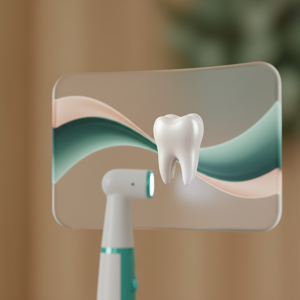
An intraoral camera is a small, pen-sized digital camera specifically designed to capture high-resolution images and video inside your mouth. The camera features a disposable protective sleeve for hygiene, LED lighting for illumination, and a lens that magnifies structures up to 25 times their actual size. When positioned near teeth or gums, the camera transmits live images to a chairside monitor, allowing you to see exactly what the dentist sees during examination.
The technology works through digital sensors that capture detailed color images of tooth surfaces, gum tissue, existing restorations, and areas of concern. Unlike trying to see your back teeth with a hand mirror, the intraoral camera provides clear, magnified views of areas impossible to see otherwise. The camera can pause on specific views, zoom in for detail, and save images for your records—creating visual documentation of your oral health status that proves invaluable for treatment planning and tracking changes over time.
Intraoral cameras fundamentally transform dental care from “trust what I tell you” to “see it for yourself.” When you can actually see a crack in your tooth, early decay beginning, or inflammation in gum tissue, treatment recommendations make immediate sense rather than seeming abstract or unnecessary. This visual understanding leads to earlier intervention, better treatment acceptance, and more consistent preventive care—all of which improve long-term oral health outcomes.
The technology also enhances diagnostic accuracy by allowing dentists to examine areas in magnified detail, catching problems at earlier, more conservative treatment stages. Small issues that might go unnoticed in standard examinations become clearly visible when magnified 10-25 times. For biological dentistry specifically, intraoral cameras help identify biofilm accumulation patterns, document biocompatibility of materials through tissue response observation, and show connections between oral inflammation and systemic health concerns. The result is more precise, personalized care based on what your specific oral environment actually shows rather than generalized assumptions.
Intraoral cameras excel at revealing early-stage problems that standard visual examination may not detect until more advanced. These include hairline cracks in teeth before they cause symptoms, beginning decay between teeth or under existing fillings, early gum recession in hard-to-see areas, worn enamel from grinding before sensitivity develops, and margin gaps in existing restorations where bacteria can penetrate. The magnification and lighting make subtle changes obvious that might otherwise progress unnoticed.
The cameras also document conditions that benefit from visual confirmation rather than verbal description. Patients often struggle to understand tongue diagnosis, tissue texture changes, or biofilm patterns without seeing them. The camera makes these observations concrete and understandable. Additionally, the technology proves valuable for monitoring healing after procedures, comparing tissue health over time, and identifying areas where home care techniques need adjustment—all through clear visual evidence rather than subjective impressions.
Intraoral camera examination typically feels completely comfortable for most patients. The camera itself is small and smooth, roughly the size of a pen, and requires no pressure or contact that would cause discomfort. The dentist or hygienist simply positions the camera near the area being examined while you rest comfortably in the chair. The entire process feels less invasive than regular dental examination with mirrors and explorers.
Some patients initially feel slight awkwardness having something in their mouth while watching the screen, but this typically passes within seconds as fascination with seeing their own teeth takes over. The examination usually takes just minutes to document your entire mouth. For patients with sensitive gag reflexes, the camera actually works better than traditional mirrors since it requires less time with instruments in the mouth and doesn’t need to reach as far back. Most patients find watching their own oral health examination interesting rather than uncomfortable—many request to see specific areas of concern in more detail.
Intraoral camera imaging transforms treatment decisions from abstract recommendations you must accept on faith to clear choices based on visual evidence you can evaluate yourself. When a dentist says “you have a cavity that needs filling,” seeing the actual decay in magnified detail makes the necessity obvious. When considering whether to address a cracked tooth now or wait, seeing the crack’s extent helps you understand the consequences of delay. This visual clarity reduces anxiety, builds confidence in treatment plans, and helps you prioritize care based on actual severity rather than guesswork.
The images also facilitate meaningful discussion about treatment options. Rather than imagining what a restoration might look like or how extensive preparation might be, you can see the existing tooth structure and understand what treatment involves. For biological dentistry decisions—such as choosing biocompatible materials, addressing mercury amalgam concerns, or understanding inflammatory patterns—the visual documentation proves invaluable. Many patients also appreciate having images to share with family members when making significant treatment decisions, or to show other healthcare providers when discussing oral-systemic health connections.
Intraoral cameras provide remarkable feedback for improving home care effectiveness. When hygienists show you areas where plaque consistently accumulates despite your brushing efforts, you can adjust technique or focus specifically where needed. Seeing biofilm patterns makes abstract flossing advice concrete—you understand exactly why those back teeth need better attention or why angled brushing matters in certain areas. This personalized, visual feedback leads to genuine improvements in home care rather than generic instructions that may not address your specific challenges.
The technology also motivates consistency by documenting improvements over time. When you can see gum tissue looking healthier, reduced inflammation, or less plaque accumulation between visits, the connection between your efforts and results becomes undeniable. For patients struggling with motivation, watching their own oral health improve through visual comparison proves far more compelling than being told “your cleaning has improved.” The camera transforms home care from a chore you should do to a practice with visible, satisfying results you can actually see.
Intraoral cameras align perfectly with biological dentistry’s emphasis on minimally invasive, biocompatible care by enabling early detection when treatment can remain conservative. Catching decay, cracks, or gum disease at early stages allows intervention before extensive tooth structure removal becomes necessary. The magnification helps identify the smallest areas of concern, supporting the biological principle of preserving maximum natural tooth structure while addressing problems before they compromise oral or systemic health.
The visual documentation also proves essential for biocompatibility assessment and mercury amalgam removal decisions. Cameras can show tissue reactions around existing materials, document inflammation patterns that may indicate material sensitivity, and provide before-after comparisons when replacing materials with biocompatible alternatives. For patients concerned about oral-systemic health connections, seeing bacterial biofilm, gum inflammation, or structural problems helps understand how oral conditions might impact overall wellness—making the mouth-body connection tangible rather than theoretical. This visual approach supports informed consent and patient autonomy, core biological dentistry values.
Yes, intraoral camera technology is standard at all Natural Dentist Associates locations throughout Rockville, Bethesda, and the DC Metro area. We consider visual documentation and patient education through direct observation essential components of quality biological dental care, not optional extras. Every operatory features chairside monitors connected to intraoral cameras, ensuring you receive the same transparent, visually-enhanced experience regardless of which location you visit.
Our commitment to this technology reflects our belief that informed patients make better health decisions and participate more actively in their care. Whether you’re visiting for routine cleaning, comprehensive evaluation, or specific treatment, you can expect your appointment to include intraoral camera documentation of relevant findings. This consistency across all locations ensures continuity of visual records and maintains our standard of care where you always understand what we’re seeing and why we’re recommending specific approaches.
All intraoral camera images captured during your appointments become part of your permanent digital dental record, stored securely in our practice management system. These images remain accessible for future appointments, allowing comparison over time to track changes, monitor healing, or document improvements. The digital storage also means images never fade, tear, or get lost like traditional film or print photographs—your visual oral health history remains available indefinitely.
Most practices can provide copies of your intraoral camera images upon request, either digitally via email or on physical media like USB drives. Having your own copies proves valuable when seeking second opinions, coordinating care with other healthcare providers, or documenting oral health for medical records. Some patients also appreciate having images to reference at home when working on technique improvements or monitoring areas of concern between visits. The images belong to your health record and accessing them should be straightforward through normal records request procedures.
Children often respond remarkably well to intraoral camera technology, finding dental examinations more interesting and less intimidating when they can watch on the screen. The visual approach transforms abstract concepts like cavities or plaque into concrete images children can understand, making oral health education far more effective. Young patients particularly enjoy seeing their own teeth “on TV” and often become more cooperative during examination when engaged by watching the camera feed.
The cameras also help children understand why certain oral health practices matter. Seeing plaque buildup makes the reason for brushing obvious. Watching tooth eruption or loose primary teeth on screen satisfies natural curiosity. For children needing treatment, seeing the problem visually reduces fear of the unknown—they understand what needs fixing and why. Parents also appreciate being able to see exactly what the dentist discusses regarding their child’s oral health, facilitating informed decisions about care. The technology turns dental visits into educational experiences rather than mysterious procedures, building positive associations with oral health care from an early age.
Intraoral cameras offer significant advantages over traditional dental photography for routine documentation and patient education. While professional dental photographs provide excellent quality for formal records, they require separate camera equipment, specific lighting setup, retractors for visibility, and post-processing time. Intraoral cameras provide instant results with minimal setup, making visual documentation practical for every examination rather than reserved for special cases.
The real-time nature of intraoral cameras also enhances communication in ways static photography cannot. You can watch as the dentist examines different areas, ask questions about specific structures as they appear on screen, and develop better spatial understanding of your oral anatomy. The dentist can demonstrate problems from multiple angles and show comparisons by moving between areas instantly. While formal dental photography certainly has its place for comprehensive documentation, intraoral cameras make visual examination and patient education practical for everyday care—transforming what was once occasional into routine practice that benefits every patient at every visit.
Intraoral camera documentation typically adds minimal time to appointments while potentially saving time overall through improved communication efficiency. Capturing and reviewing key images usually takes just 2-3 minutes, and this investment often eliminates extended verbal explanations that patients might not fully understand without visual reference. When you can see a problem clearly, explanations become brief and focused rather than lengthy attempts to help you imagine what the dentist describes.
The technology may actually shorten total treatment time by reducing misunderstandings and treatment hesitation. Patients who clearly see and understand problems tend to accept necessary treatment more readily rather than postponing due to uncertainty. The visual documentation also reduces time spent at future appointments explaining previous findings or treatment rationale—the images provide instant context. For comprehensive examinations or consultations where thorough documentation benefits treatment planning, the camera proves invaluable. For routine cleanings, camera use typically remains brief and focused. Overall, most patients feel the slight time addition provides substantial value through better understanding and confidence in their care.
During your first intraoral camera examination, expect a fascinating perspective on your oral health you’ve likely never experienced. The dentist or hygienist will explain the technology briefly, then begin systematically documenting your mouth while you watch the live feed on a chairside monitor. You’ll see your teeth magnified far beyond normal vision, making details like enamel texture, existing restoration margins, and tissue characteristics clearly visible. Most patients find this initial viewing surprising—their teeth look quite different up close than imagined.
The clinician will likely pause on areas of interest to explain what you’re seeing, point out healthy structures for reference, and discuss any concerns. Feel free to ask questions about anything that catches your attention or request closer looks at specific areas. The examination remains comfortable throughout, with the camera moving smoothly from area to area without requiring you to hold positions for extended periods. By the end of your first camera-enhanced examination, you’ll likely have dramatically better understanding of your oral health status and greater confidence in recommended care—most patients wish they’d experienced this visual approach years earlier.
Ready to See Your Dental Health Clearly?
Contact Natural Dentist Associates today to experience dentistry where you see exactly what we see, understand every recommendation, and participate actively in your oral health journey. Our intraoral camera technology transforms dental visits from mysterious to transparent.